PATHOLOGICAL FEATURES OF SPERMATOCYTIC SEMINOMA IN CARASSIUS GIBELIO
-
摘要: 自银鲫(Carassius gibelio)中发现了一例自发性精母细胞型精原细胞瘤, 详细描述了该肿瘤的组织病理和超微结构。在组织学上, 该肿瘤具有薄膜包被、扩张性生长, 具有高度细胞性和弥漫性结缔组织, 伴有淋巴细胞浸润。透射电镜显示, 该肿瘤显著的特征是含有未分化的精母细胞, 且支撑细胞稀少。对比正常银鲫的精巢组织结构, 排除了其他肿瘤类型, 最终将该肿瘤鉴定为精母细胞型精原细胞瘤。系在低等脊椎动物中首次发现具有淋巴细胞浸润的精母细胞型精原细胞瘤, 丰富了鱼类性腺肿瘤的基础资料, 也为构建人类精母细胞型精原细胞瘤动物模型提供了依据。
-
关键词:
- 银鲫 /
- 肿瘤 /
- 精母细胞型精原细胞瘤 /
- 组织结构
Abstract: This study described pathological features of a spontaneous spermatocytic seminoma in gibel carp Carassius gibelio. Histologically, the neoplasm founded in the abdomen was encapsulated invasive spermatocytic seminoma with an inconspicuous lobular architecture and diffuse fibro-connective septa with lymphocytic infiltration. For its ultrastructure, undifferentiated spermatocytes and fewer supporting cells were the unique features of the neoplasm. Compared with the histological features of normal testis of gibel carp, this neoplasm was identified as a spermatocytic seminoma. The first report of spermatocytic seminoma with lymphocytic infiltration in fish may contribute to the establishment of fish model of spermatocytic seminoma.-
Keywords:
- Carassius gibelio /
- Tumor /
- Spermatocytic seminoma /
- Histopathology
-
肿瘤是机体在各种致瘤因素作用下, 局部组织的细胞基因调控失常, 导致克隆性异常增生而形成的局部肿块, 是当前人类健康的主要威胁之一[1]。良性肿瘤对器官、组织只有挤压和阻塞作用, 易被清除干净, 一般不转移、不复发。恶性肿瘤能破坏组织、器官的结构和功能, 引起坏死、出血及感染等, 并通过血液及淋巴系统转移形成转移灶, 导致多脏器的损害[2]。与高等脊椎动物类似, 鱼类也发现了多种肿瘤。尽管鱼类肿瘤在养殖中的危害较轻, 但作为肿瘤研究的工具, 已成为人类肿瘤的发生、发展等机理研究以及抗癌药物筛选不可或缺的模型。鱼类肿瘤还作为环境指示物在致癌物鉴定等领域发挥重要作用。
至今, 已记录的鱼类肿瘤包括174例, 分属于35种, 包括乳头状肿瘤、神经胚胎瘤、肾胚胎瘤、黑色素瘤、纤维瘤、性腺肿瘤等, 其中皮肤瘤报道最多, 而性腺肿瘤较少[3—5]。鱼类性腺肿瘤的报道仅有12例, 分别发生在鲤科、肺鱼科、太阳鱼科和鮨科等4科7种[3—6]。鱼类性腺肿瘤主要有以下三类: 一是自发性的性腺基质肿瘤, 如支持细胞肿瘤, 仅在鲤科鱼类中发现[6]; 二是间质细胞性腺肿瘤, 如平滑肌瘤和纤维肌瘤, 在黄鲈、大口黑鲈和非洲肺鱼等鱼中发现[7—9]; 三是生殖细胞肿瘤, 如典型精原细胞瘤和精母细胞型精原细胞瘤, 已在鲤科、肺鱼科鱼类中发现[6, 10, 11] 。
精原细胞瘤也称生殖细胞癌, 在生殖细胞肿瘤中约占60%, 高发于雄性动物[4]。典型精原细胞瘤原发于生殖腺, 在睾丸处较常见, 也可见于后腹膜、前列腺、肺、胸腺、外耳道等部位。精母细胞型精原细胞瘤约占生殖细胞肿瘤的4%—7%, 常发于年龄较大的雄性, 一般不发生转移, 但具有淋巴细胞浸润的精母细胞型精原细胞瘤较罕见[12]。本病的病因尚不明确, 可能与遗传、隐睾、内分泌、外伤有关, 此外病毒性疾病和细菌性感染也可间接引发性腺肿瘤。鱼类的精原细胞瘤分别在金鱼和鲈中发现, 并已建立斑马鱼模型[13—15],但精母细胞型精原细胞瘤鱼类模型尚未建立。
本研究报道了一例银鲫(Carassius gibelio)自发性精母细胞型精原细胞瘤, 详细描述了其病理特征及其主要细胞类型, 系在低等脊椎动物中首次发现具有淋巴细胞浸润的精母细胞型精原细胞瘤, 丰富了鱼类性腺肿瘤的研究资料, 也为构建人类精母细胞型精原细胞瘤动物模型提供了依据。
1. 材料与方法
1.1 材料
银鲫采集自某湖泊, 经充氧运输至实验室, 用于实验教学。解剖过程中发现一例患肿瘤的银鲫, 分离肿瘤组织后用于后续的分析。
1.2 组织切片制备及HE染色
将摘除的肿瘤组织, 切成0.5 cm×0.5 cm小块, 4%多聚甲醛固定24h。石蜡包埋后, 连续切片(制备组织3 μm切片)。烘干后依次经乙醇梯度脱水、二甲苯透明, HE染色封片[16]。按相同方法取健康鲫的精巢制片, 在OLYMPUS CX21显微镜下观察并拍照。
1.3 超薄切片制备与观察
自摘除的肿瘤组织, 切成3 mm×3 mm小块, 以戊二醛固定48h, 超薄切片由中国科学院武汉病毒研究所协助完成, 制备的切片在200 KV透射电镜下(TECNAL, FEI)观察并拍照。
2. 结果
2.1 大体观察
宏观上, 鱼体明显消瘦, 腹部肿大, 触摸有硬块, 鳞片和鳍均正常。解剖发现该鱼体内腹腔后部紧邻泄殖孔处有一被膜完整的实质肿块, 呈淡黄色, 大小为3 cm×2 cm×1.5 cm (图 1a)。肿块横切面坚实, 质地均匀。对肿块的边界检查证实了它们的限制性, 未见向邻近组织如肠壁、肝胰脏、脾脏、肾脏和鳔浸润(图 1b)。
2.2 组织结构观察
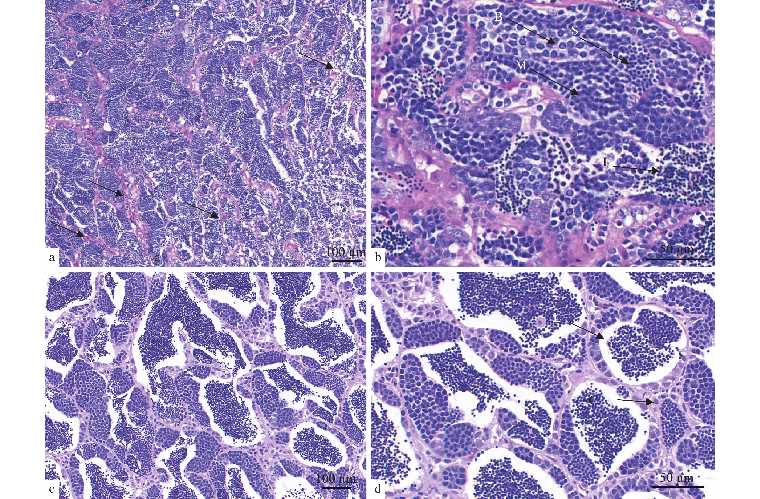
肿瘤组织经制备石蜡切片后HE染色, 显微观察显示, 肿瘤组织表现扩张性生长和血管增生, 肿瘤间质组织弥漫排列, 肿瘤细胞排列较密, 几乎充满整个精小巢(图 2a)。瘤细胞大小不一, 主要包括大、中、小三种细胞类型, 形状为圆形或椭圆形。细胞核位于中央, 核呈圆形或椭圆形, 可见小的核仁, 有的胞浆呈现空泡状。部分区域有裂隙, 裂隙边缘被覆肿瘤细胞(图 2b)。肿瘤间质组织呈高度细胞性和弥散性, 缺乏间质细胞, 主要由纤维成分组成, 其中小血管较为常见(图 2a、2b)。肿瘤组织中可见淋巴细胞浸润, 表现为轻度的炎症反应。对比正常的精巢结构(图 2c、2d), 其为多细胞巢, 结缔组织伸入精巢内部形成许多隔膜, 具有明显的小叶结构特征。
![]() 图 2 精母细胞型精原细胞瘤的组织结构(HE)a、b. 肿瘤组织的精巢结构: 肿瘤组织具有高度细胞性和弥漫性的间质组织; a. 肿瘤组织具有血管增生(↑); b. 肿瘤组织主要包含三种细胞类型, 大细胞(B)、中等细胞(M)、小细胞(S), 肿瘤细胞形态类似于初级精母细胞, 肿瘤组织具有轻度的淋巴细胞浸润(I); c、d. 正常的精巢呈小叶状结构(L), 结缔组织伸入精巢内部形成许多隔膜(C), 把精巢分成许多精小叶Figure 2. Histological structure of spermatocytic seminomaa, b. Tumor tissue. The tumor was diffused by fibro-connective septa and cells, Dyeing: hematoxylin-eosin (HE); a. Tumor angiogenesis (↑). b. Neoplasm mainly contains large cells (D), medium cells (M), and small cells (S). The morphology of tumor cells are similar to the primary spermatocyte and the tumor tissue have a mild lymphocyte infiltration (☆); c, d. The normal testis shows a lobular structure (L). Connective tissue extends into the nest to form a number of diaphragm (C) and divides the nest into many fine leaves. Dyeing: HE
图 2 精母细胞型精原细胞瘤的组织结构(HE)a、b. 肿瘤组织的精巢结构: 肿瘤组织具有高度细胞性和弥漫性的间质组织; a. 肿瘤组织具有血管增生(↑); b. 肿瘤组织主要包含三种细胞类型, 大细胞(B)、中等细胞(M)、小细胞(S), 肿瘤细胞形态类似于初级精母细胞, 肿瘤组织具有轻度的淋巴细胞浸润(I); c、d. 正常的精巢呈小叶状结构(L), 结缔组织伸入精巢内部形成许多隔膜(C), 把精巢分成许多精小叶Figure 2. Histological structure of spermatocytic seminomaa, b. Tumor tissue. The tumor was diffused by fibro-connective septa and cells, Dyeing: hematoxylin-eosin (HE); a. Tumor angiogenesis (↑). b. Neoplasm mainly contains large cells (D), medium cells (M), and small cells (S). The morphology of tumor cells are similar to the primary spermatocyte and the tumor tissue have a mild lymphocyte infiltration (☆); c, d. The normal testis shows a lobular structure (L). Connective tissue extends into the nest to form a number of diaphragm (C) and divides the nest into many fine leaves. Dyeing: HE2.3 超微结构观察
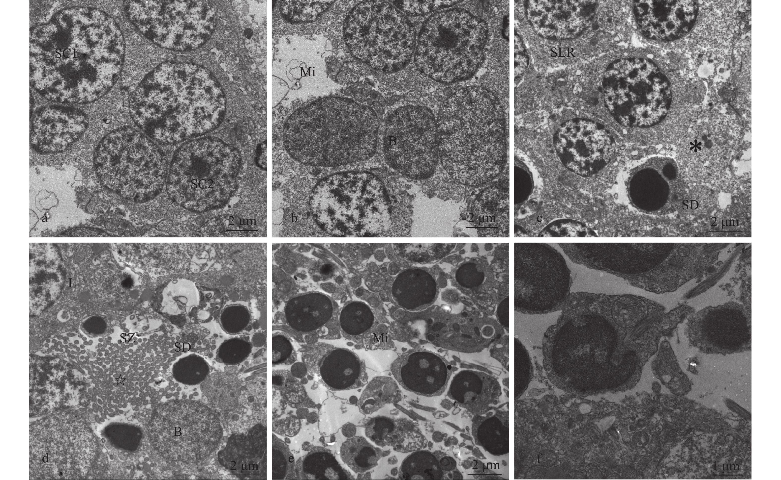
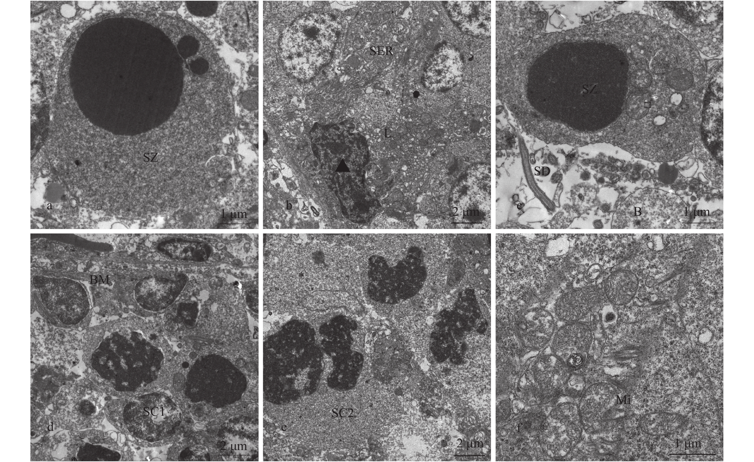
透射电镜观察显示: 肿瘤组织中初级精母细胞(SC1)是主要的细胞类型, 次级精母细胞(SC2)比初级精母细胞(SC1)小, 但形态相似。精母细胞之间缺乏桥粒特征(图 3a—c)。该肿瘤组织最显著的特征是大部分细胞停留在精母细胞阶段(图 3a、3b), 且精细胞凋亡(图 3c)。精细胞(SD)在精子发生完成之前释放包含在巢中, 缺乏支撑细胞连接(图 3e、3f), 其中一些精细胞凋亡和变性(图 4a、4b)。间质组织含有很少的支撑细胞, 这些支撑细胞位于生精小叶的周边, 与基膜接触(图 4c)。间质组织中含有电子致密脂滴, 部分空泡化(图 4d)。另外细胞质含有大量的游离核糖体和少量的膜结合颗粒(图 4e)。基于发现未分化的生殖细胞和少量的有丝分裂(图 4d—f), 据此将该肿瘤病变分类为精母细胞型精原细胞瘤。
![]() 图 3 精母细胞型精原细胞瘤的超微结构a、b. SC1细胞是主要的细胞类型, 形成大的细胞巢, 含有大量的线粒体, 但缺乏桥粒特征; c. 细胞巢内的精细胞; d. 未完全发育精细胞被包含在巢中, 缺乏支持细胞连接。“☆”精子尾管结构; e、f. 精子细胞被释放到基质的边缘或进入管腔内; f.未分化的精细胞; SC1: 初级精母细胞; SC2: 次级精母细胞; SD: 精细胞; SZ: 精子; Mi: 线粒体; B: B型精原细胞; SER: 滑面内质网; L: 脂滴; *: 支持细胞Figure 3. Ultrastructure of spermatocytic seminomaa, b. SC1 cells are the major cell types and form large cell nests. SC1 cells contain a large number of mitochondria, but lack bridge particles; c. Sperm cells was in cell nests; d. Spermatid was encompassed in the nest and not connected with Sertoli cell. “*”: sperm tail pipe structure; e, f. Sperm cells are released to the edge of the substrate or into the lumen; f. Undifferentiated spermatid; SC1: primary spermatocyte; SC2: secondary spermatocyte; SD: spermatid; SZ: spermatozoa; Mi: mitochondria; B: type B spermatophore; SER: smooth endoplasmic reticulum; L: lipid drops; *: supporting cells
图 3 精母细胞型精原细胞瘤的超微结构a、b. SC1细胞是主要的细胞类型, 形成大的细胞巢, 含有大量的线粒体, 但缺乏桥粒特征; c. 细胞巢内的精细胞; d. 未完全发育精细胞被包含在巢中, 缺乏支持细胞连接。“☆”精子尾管结构; e、f. 精子细胞被释放到基质的边缘或进入管腔内; f.未分化的精细胞; SC1: 初级精母细胞; SC2: 次级精母细胞; SD: 精细胞; SZ: 精子; Mi: 线粒体; B: B型精原细胞; SER: 滑面内质网; L: 脂滴; *: 支持细胞Figure 3. Ultrastructure of spermatocytic seminomaa, b. SC1 cells are the major cell types and form large cell nests. SC1 cells contain a large number of mitochondria, but lack bridge particles; c. Sperm cells was in cell nests; d. Spermatid was encompassed in the nest and not connected with Sertoli cell. “*”: sperm tail pipe structure; e, f. Sperm cells are released to the edge of the substrate or into the lumen; f. Undifferentiated spermatid; SC1: primary spermatocyte; SC2: secondary spermatocyte; SD: spermatid; SZ: spermatozoa; Mi: mitochondria; B: type B spermatophore; SER: smooth endoplasmic reticulum; L: lipid drops; *: supporting cells![]() 图 4 精母细胞型精原细胞瘤的超微结构a、b. 精子细胞凋亡和变性; c. 精子细胞含有许多线粒体、溶酶体和致密的脂滴, 线粒体形状也发生了变化; d. 分化不良的生殖细胞; e. 初级精母细胞的有丝分裂; f. 肿瘤组织的间质组织包含少量支持细胞和大量细胞器; SC1: 初级精母细胞; SC2: 次级精母细胞; SD: 精细胞; SZ: 精子; Mi: 线粒体; B: B型精原细胞; SER: 滑面内质网; L: 脂滴; BM: 基膜; *: 支持细胞; □: 近端中心粒; ▲: 间质细胞Figure 4. Ultrastructure of spermatocytic seminomaa, b. Apoptosis and degeneration of sperm cell; c. Spermatid containing many mitochondrial lysosomes and dense lipid droplets as well as abnormal mitochondria. d. Germ cells had poor differentiation; e. Primary spermatocytes in mitotic; f. Mesenchymal tissue of tumor containing a small number of supporting cells with plentiful organelles; SC1: primary spermatocyte; SC2: secondary spermatocyte; SD: spermatid; SZ: spermatozoa; Mi: mitochondria; B: type B spermatophore; SER: smooth endoplasmic reticulum; L: lipid drops; BM: basement membrane; *: supporting cells; □: proximal centrioles; ▲: Interstitial cells
图 4 精母细胞型精原细胞瘤的超微结构a、b. 精子细胞凋亡和变性; c. 精子细胞含有许多线粒体、溶酶体和致密的脂滴, 线粒体形状也发生了变化; d. 分化不良的生殖细胞; e. 初级精母细胞的有丝分裂; f. 肿瘤组织的间质组织包含少量支持细胞和大量细胞器; SC1: 初级精母细胞; SC2: 次级精母细胞; SD: 精细胞; SZ: 精子; Mi: 线粒体; B: B型精原细胞; SER: 滑面内质网; L: 脂滴; BM: 基膜; *: 支持细胞; □: 近端中心粒; ▲: 间质细胞Figure 4. Ultrastructure of spermatocytic seminomaa, b. Apoptosis and degeneration of sperm cell; c. Spermatid containing many mitochondrial lysosomes and dense lipid droplets as well as abnormal mitochondria. d. Germ cells had poor differentiation; e. Primary spermatocytes in mitotic; f. Mesenchymal tissue of tumor containing a small number of supporting cells with plentiful organelles; SC1: primary spermatocyte; SC2: secondary spermatocyte; SD: spermatid; SZ: spermatozoa; Mi: mitochondria; B: type B spermatophore; SER: smooth endoplasmic reticulum; L: lipid drops; BM: basement membrane; *: supporting cells; □: proximal centrioles; ▲: Interstitial cells3. 讨论
2016年世界卫生组织对人类和哺乳动物睾丸肿瘤进行了新分类, 根据临床病例特点将睾丸肿瘤分为两个亚型, 精母细胞肿瘤和精原细胞瘤[17]。精原细胞瘤主要发生在睾丸及性腺外, 伴随其他生殖细胞肿瘤, 细胞类型单一且纤维性间质显著; 精母细胞瘤主要发生于睾丸, 不伴随其他类型的生殖细胞瘤, 细胞类型有大、中、小三种细胞且纤维性间质稀少。Masahito等[11]报道的人精母细胞瘤主要由大细胞、中等细胞、小细胞构成, 缺乏糖原及肉芽肿反应、间质少, 肿瘤细胞形态类似于初级精母细胞。在本研究中银鲫的肿瘤组织中有含大量的未完全成型精细胞, 且存在精子的尾管结构, 排除了肝癌、肠癌等其他肿瘤的干扰, 证实为精原细胞瘤。超微结果观察发现, 肿瘤组织内的细胞主要包括大细胞、中等细胞和小细胞构成, 支撑细胞稀少, 与人类和哺乳动物的精母细胞瘤一致, 因此将该性腺肿瘤定义为精母细胞型精原细胞瘤。
同已报道的鱼类精原细胞瘤比较, 本研究描述的银鲫精原细胞瘤具有不同发育阶段的精母细胞, 且可维持精子的正常发育阶段, 与Nigrelli和Jakowska[18]和Masahito等[11]所描述的精母细胞型精原细胞瘤一致, 但这些肿瘤组织不具有淋巴细胞的浸润。我们在本例精原细胞瘤组织中还观察到较少的有丝分裂现象和稀少的支撑细胞, 并有少量的淋巴细胞浸润, 这与Hubbard和Fletcher[10]中所描述的病理特征差别较大, 该精原细胞瘤由分化较好的大肿瘤细胞群组成, 有丝分裂率高, 没有明显的炎症和坏死。Hawkins 等[19]和Smolowitz 等[13]描述的精原细胞瘤病理特征是具有被丰富的纤维结缔组织分隔小叶模式, 精子细胞聚集, 具有低有丝分裂率的肿瘤细胞, 而本例精母型精原细胞瘤具有高度细胞性和弥散的结缔组织, 缺乏明显的小叶结构。
根据世界卫生组织的家畜生殖系统肿瘤国际组织学分类, 精原细胞瘤可分为内瘤型和弥漫型2种生长模式[20]。本例肿瘤的显著特点为肿瘤细胞弥漫分布, 大小不一, 支持细胞少, 未侵入其他组织。肿瘤细胞的小叶排列和由淋巴细胞浸润的基质成分, 表明本例精原细胞瘤为弥漫型。鱼类的性腺肿瘤一般表现为低度恶性, 具有低度分化的肿瘤细胞, 肿瘤组织有高度的胞核细胞和异核细胞, 有的案例肿瘤组织侵犯临近组织, 有的案例肿瘤组织被完整的包膜包裹, 不浸润临近器官。在人类和其他哺乳动物中, 睾丸生殖细胞来源的多数精原细胞瘤也为低度恶性肿瘤, 如犬睾丸精原细胞瘤[21]。本研究报道的精母细胞型精原细胞瘤, 宏观上具包膜包被、实质, 呈扩大性生长, 压迫肠道和肝脏组织, 生殖腺可辨认, 推断该精原细胞瘤起源于非正常位置。
鱼类肿瘤作为肿瘤学研究的工具已取得较大进展。新月鱼和剑尾鱼的杂交一代自发产生的黑色素瘤已经成为研究肿瘤形成的重要模型[22]; 双色雀鲷中由神经纤维瘤和色素细胞瘤组成的多发性神经纤维瘤疾病, 已作为人类罕见的多发性神经纤维瘤 1型疾病的研究模型[23], 且已经成功建立虹鳟肝细胞瘤和肾母细胞瘤模型、金鱼肝肿瘤和神经鞘瘤模型、鲴乳头状瘤模型、白斑狗鱼淋巴瘤模型等[24]。鱼类精原细胞瘤与哺乳动物的精原细胞瘤在形态结构上具有一致性。Sirri等[6]的肿瘤免疫组化表型也证明了人类、哺乳动物和鱼类性腺肿瘤的相似性。目前, 针对于精母型精原细胞瘤的研究较少, 本病例的发现为建立此肿瘤的鱼类模型提供了参考。
尽管诱发肿瘤的因素复杂, 但环境因素是最为重要的诱因之一[3]。1975年, Gennady等[25]用N-2-氟乙烯乙酰胺、O-氨基偶氮甲苯、4-二甲氨基偶氮苯、二乙基亚硝胺和二甲基亚硝胺等成功诱导了水族箱中孔雀鱼和斑马鱼的胆管瘤、肝腺瘤、胆管癌和肝细胞癌等, 证明了鱼类是检测致癌化合物和筛选环境致癌物质的合适模型。陈玉汉等[26]1966—1971年的流行病学调查显示鱼类精原细胞瘤五年平均检出率为10.8%, 类似现象在西班牙塔古斯河、水库和盆地中也观察到[27, 28]。而Down和Leatherland [29]发现鱼类肿瘤在工业污染较严重或者是相对不受工业污染的地区都有存在。因此, 通过监测水体内鱼类的肿瘤检出率, 能否作为环境致癌物的评估指标尚需更全面的调查。
-
图 2 精母细胞型精原细胞瘤的组织结构(HE)
a、b. 肿瘤组织的精巢结构: 肿瘤组织具有高度细胞性和弥漫性的间质组织; a. 肿瘤组织具有血管增生(↑); b. 肿瘤组织主要包含三种细胞类型, 大细胞(B)、中等细胞(M)、小细胞(S), 肿瘤细胞形态类似于初级精母细胞, 肿瘤组织具有轻度的淋巴细胞浸润(I); c、d. 正常的精巢呈小叶状结构(L), 结缔组织伸入精巢内部形成许多隔膜(C), 把精巢分成许多精小叶
Figure 2. Histological structure of spermatocytic seminoma
a, b. Tumor tissue. The tumor was diffused by fibro-connective septa and cells, Dyeing: hematoxylin-eosin (HE); a. Tumor angiogenesis (↑). b. Neoplasm mainly contains large cells (D), medium cells (M), and small cells (S). The morphology of tumor cells are similar to the primary spermatocyte and the tumor tissue have a mild lymphocyte infiltration (☆); c, d. The normal testis shows a lobular structure (L). Connective tissue extends into the nest to form a number of diaphragm (C) and divides the nest into many fine leaves. Dyeing: HE
图 3 精母细胞型精原细胞瘤的超微结构
a、b. SC1细胞是主要的细胞类型, 形成大的细胞巢, 含有大量的线粒体, 但缺乏桥粒特征; c. 细胞巢内的精细胞; d. 未完全发育精细胞被包含在巢中, 缺乏支持细胞连接。“☆”精子尾管结构; e、f. 精子细胞被释放到基质的边缘或进入管腔内; f.未分化的精细胞; SC1: 初级精母细胞; SC2: 次级精母细胞; SD: 精细胞; SZ: 精子; Mi: 线粒体; B: B型精原细胞; SER: 滑面内质网; L: 脂滴; *: 支持细胞
Figure 3. Ultrastructure of spermatocytic seminoma
a, b. SC1 cells are the major cell types and form large cell nests. SC1 cells contain a large number of mitochondria, but lack bridge particles; c. Sperm cells was in cell nests; d. Spermatid was encompassed in the nest and not connected with Sertoli cell. “*”: sperm tail pipe structure; e, f. Sperm cells are released to the edge of the substrate or into the lumen; f. Undifferentiated spermatid; SC1: primary spermatocyte; SC2: secondary spermatocyte; SD: spermatid; SZ: spermatozoa; Mi: mitochondria; B: type B spermatophore; SER: smooth endoplasmic reticulum; L: lipid drops; *: supporting cells
图 4 精母细胞型精原细胞瘤的超微结构
a、b. 精子细胞凋亡和变性; c. 精子细胞含有许多线粒体、溶酶体和致密的脂滴, 线粒体形状也发生了变化; d. 分化不良的生殖细胞; e. 初级精母细胞的有丝分裂; f. 肿瘤组织的间质组织包含少量支持细胞和大量细胞器; SC1: 初级精母细胞; SC2: 次级精母细胞; SD: 精细胞; SZ: 精子; Mi: 线粒体; B: B型精原细胞; SER: 滑面内质网; L: 脂滴; BM: 基膜; *: 支持细胞; □: 近端中心粒; ▲: 间质细胞
Figure 4. Ultrastructure of spermatocytic seminoma
a, b. Apoptosis and degeneration of sperm cell; c. Spermatid containing many mitochondrial lysosomes and dense lipid droplets as well as abnormal mitochondria. d. Germ cells had poor differentiation; e. Primary spermatocytes in mitotic; f. Mesenchymal tissue of tumor containing a small number of supporting cells with plentiful organelles; SC1: primary spermatocyte; SC2: secondary spermatocyte; SD: spermatid; SZ: spermatozoa; Mi: mitochondria; B: type B spermatophore; SER: smooth endoplasmic reticulum; L: lipid drops; BM: basement membrane; *: supporting cells; □: proximal centrioles; ▲: Interstitial cells
-
[1] 徐晓艳, 夏养志, 徐晓锋. 肿瘤侵袭、转移的亚临床研究. 山西医科大学学报, 2003, 34(4): 360—363 doi: 10.3969/j.issn.2095-1450.2003.04.018 Xu X Y, Xia Y Z, Xu X F. A subclinical study of tumor invasion and metastasis [J]. Journal of Shanxi Medical University, 2003, 34(4): 360—363 doi: 10.3969/j.issn.2095-1450.2003.04.018
[2] 刘彤华. 内分泌肿瘤组织病理学及分子病理学研究进展. 中华病理学杂志, 2004, 33(3): 193—194 doi: 10.3760/j.issn:0529-5807.2004.03.001 Liu T H. Research progress of endocrine tumor histopathology and molecular pathology [J]. Chinese Journal of Pathology, 2004, 33(3): 193—194 doi: 10.3760/j.issn:0529-5807.2004.03.001
[3] Groff J M. Neoplasia in fishes [J]. Veterinary Clinics of North America Exotic Animal Practice, 2004, 7(3): 705—756 doi: 10.1016/j.cvex.2004.04.012
[4] Harshbarger J C. Chronology of oncolygy in fish, amphibians and invertebrates [J]. Bulletin-European Association of Fish Pathologists, 2004, 24(1): 62—84
[5] Palikova M, Navratil S, Svobodova Z, et al. Skin and gonadal tumours in a barbel (Barbus barbus) – a case report [J]. Bulletin-European Association of Fish Pathologists, 2007, 27(6): 234—238
[6] Sirri R, Mandrioli L, Grieco V, et al. Seminoma in a koi carp Cyprinus carpio: histopathological and immunohistochemical findings [J]. Diseases of Aquatic Organisms, 2010, 92(1): 83—88 doi: 10.3354/dao02273
[7] Herman R L, Landolt M. A testicular leiomyoma in a largemouth bass, Micropterus salmoides [J]. Journal of Wildlife Diseases, 1975, 11(1): 128 doi: 10.7589/0090-3558-11.1.128
[8] Blazer V S. Histopathological assessment of gonadal tissue in wild fishes [J]. Fish Physiology & Biochemistry, 2002, 26(1): 85—101
[9] Weisse C, Weber E S, Matzkin Z, et al. Surgical removal of a seminoma from a black sea bass [J]. Journal of the American Veterinary Medical Association, 2002, 221(2): 280 doi: 10.2460/javma.2002.221.issue-2
[10] Hubbard G B, Fletcher K C. A seminoma and a leiomyosarcoma in an Albino African Lungfish (Protopterus dolloi) [J]. Journal of Wildlife Diseases, 1985, 21(1): 72—74 doi: 10.7589/0090-3558-21.1.72
[11] Masahito P, Ishikawa T, Takayama S. Spontaneous spermatocytic seminoma in African lungfish, Protopterus aethiopicus Heckel [J]. Journal of Fish Diseases, 2010, 7(2): 169—172
[12] 詹玛琍, 李传应, 顾萍, 等. 睾丸精原细胞瘤临床病理特征与免疫表型分析. 解剖学研究, 2016, (6): 437—440 Zhan M L, Li C Y, Gu P, et al. The clinicopathological features and immunophenotypic analysis [J]. Anatomical Studies, 2016, (6): 437—440
[13] Smolowitz R, Hanley J H. A three-year retrospective study of abdominal tumors in zebrafish maintained in an aquatic laboratory animal facility [J]. Biological Bulletin, 2002, 203(2): 265 doi: 10.2307/1543433
[14] Basten S G, Davis E E, Gillis A J, et al. Mutations in LRRC50 predispose zebrafish and humans to seminomas [J]. PLoS Genetics, 2013, 9(4): e1003384 doi: 10.1371/journal.pgen.1003384
[15] Spitsbergen J M, Kent M L. The state of the art of the zebrafish model for toxicology and toxicologic pathology research—advantages and current limitations [J]. Toxicologic Pathology, 2003, 31Suppl(1): 62
[16] 黄灿, 张忠海, 吴淑勤, 等. 益生芽孢杆菌对草鱼肠黏膜结构的保护作用. 水生生物学报, 2017, 41(4): 774—780 Huang Can, Zhang Z H, Wu S Q, et al. The protective effect of probiotic Bacillus on the intestinal mucosal structure of Ctenopharyngodon indllus [J]. Acta Hydrobiologica Sinica, 2017, 41(4): 774—780
[17] 吕炳建, 程亮. 2016版WHO睾丸和阴茎肿瘤新分类解读. 中华病理学杂志, 2016, 45(8): 518—521 doi: 10.3760/cma.j.issn.0529-5807.2016.08.004 Lü B J, Cheng J. New classification interpretation of the WHO testis and penile tumors in 2016 [J]. Chinese Journal of Pathology, 2016, 45(8): 518—521 doi: 10.3760/cma.j.issn.0529-5807.2016.08.004
[18] Nigrelli RF, Jakowska S. Spontaneous neoplasms in fishes, Vol 7. A spermatocytoma and renal melanoma in an African lungfish, Protopterus anneetens (Owen) [J]. Zoologica, 1953, 38: 109—112
[19] Hawkins W E, Fournie J W, Ishikawa T, et al. Germ cell neoplasms in Japanese Medaka [J]. Journal of Aquatic Animal Health, 1996, 8(2): 120—129 doi: 10.1577/1548-8667(1996)008<0120:GCNIJM>2.3.CO;2
[20] Kennedy P C. Histological Classification of Tumors of the Genital System of Domestic Animals [M]. Armed Forces Institute of Pathology. 1998, 17
[21] 韩庆功, 刘世忠, 耿莎莎, 等. 犬睾丸精原细胞瘤病理学观察与分析. 畜牧与兽医, 2017, 49(10): 46—49 Han Q G, Liu S Z, Geng S S, et al. Pathological observation and analysis of spermatogenic cell tumor in dogs [J]. Animal Husbandry and Veterinary Medicine, 2017, 49(10): 46—49
[22] 李凯彬, 常藕琴, 石存斌, 等. 剑尾鱼杂交种黑色素瘤的形成和病理观察. 中国实验动物学报, 2005, 13(2): 76—78 doi: 10.3969/j.issn.1005-4847.2005.02.004 Li K B, Chang O Q, Shi C B, et al. Formation and pathological observation of melanoma of swordtail hybrids [J]. Chinese Journal of Experimental Animals, 2005, 13(2): 76—78 doi: 10.3969/j.issn.1005-4847.2005.02.004
[23] 江千秋, 刘冬娥, 田智, 等. 鱼类实验动物的生物学特性及应用. 湖南生态科学学报, 2006, 12(2): 129—132 doi: 10.3969/j.issn.1671-6361.2006.02.004 Jiang Q Q, Liu D E, Tian Z, et al. Biological characteristics and application of fish experimental animals [J]. Journal of Hunan Ecological Science, 2006, 12(2): 129—132 doi: 10.3969/j.issn.1671-6361.2006.02.004
[24] 吴端生, 王宗保. 鱼类实验动物开发与应用研究的现状及展望. 中国比较医学杂志, 2000, 10(2): 103—109 doi: 10.3969/j.issn.1671-7856.2000.02.010 Wu D S, Wang Z B. The present situation and prospect of the development and application of fish experimental animals [J]. Journal of Comparative Medicine in China, 2000, 10(2): 103—109 doi: 10.3969/j.issn.1671-7856.2000.02.010
[25] Pliss G B, Khudoley V V. Tumor induction by carcinogenic agents in aquarium fish [J]. Journal of the National Cancer Institute, 1975, 55(1): 129—136 doi: 10.1093/jnci/55.1.129
[26] 陈玉汉, 陈灼怀, 肖振德. 鱼类肿瘤. 广东科技出版社. 1993, 107—108 Chen Y H, Chen Z H, Xiao Z D. Fish Tumor [M]. Guangdong Science and Technology Press. 1993, 107—108
[27] Dickman M D, Steele P O. Gonadal neoplasms in wild carp-goldfish hybrids from the Welland River near Niagara Falls, Canada [J]. Hydrobiologia, 1986, 134(3): 257—263 doi: 10.1007/BF00008494
[28] Leatherland J F, Sonstegard R A. Structure of normal testis and testicular tumors in cyprinids from Lake Ontario [J]. Cancer Research, 1978, 38(10): 3164
[29] Down N E, Leatherland J F. Histopathology of gonadal neoplasms in cyprinid fish from the lower Great Lakes of North America [J]. Journal of Fish Diseases, 1989, 12(5): 415—437 doi: 10.1111/jfd.1989.12.issue-5


 下载:
下载:


